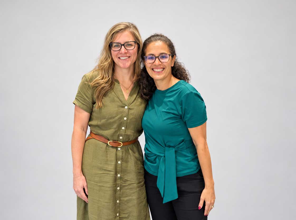

By: Keahealailani Takushi-Coffey, DO
Editor’s Note: Keahealailani Takushi-Coffey, DO, wrote this article during her fourth year of medical school while participating in the FACTS elective. She examined research by Elizabeth J. Houston and Nia M. Templeman that challenges the traditional understanding of PMOS by proposing that hyperinsulinemia may precede—and contribute to—insulin resistance, with important implications for ovarian function and fertility. In recognition of PMOS Awareness Month, this article highlights how fertility awareness-based methods (FABMs) can help women better understand irregular cycles and other biomarkers associated with PMOS while offering clinicians valuable insight into each patient’s hormonal and metabolic health.
Polycystic ovary syndrome (PCOS), now renamed polyendocrine metabolic ovarian syndrome (PMOS), is the most common endocrine disorder among women of reproductive age and is a significant cause of ovulatory infertility [1],[2]. Insulin resistance and hyperinsulinemia are present in approximately 60–95% of patients with PMOS, and it plays a central role in perpetuating hyperandrogenism and ovarian dysfunction [3] [4] [5]. Traditional literature has suggested that PMOS is primarily driven by insulin resistance, with compensatory hyperinsulinemia developing as a secondary consequence that contributes to downstream reproductive and metabolic dysfunction [6]. However, “Reappraising the Relationship Between Hyperinsulinemia and Insulin Resistance in PCOS” challenges this paradigm by proposing that hyperinsulinemia itself may be the primary abnormality in PMOS, with insulin resistance developing later as a protective physiologic response [7]. Reframing the cause-and-effect relationship between insulin resistance and hyperinsulinemia has important implications for understanding PMOS physiology, particularly in the context of fertility awareness and fertility awareness–based methods (FABMs), which rely on recognizing and interpreting hormonal patterns within the female reproductive cycle.
“Insulin resistance and hyperinsulinemia are present in approximately 60–95% of patients with PMOS, and it plays a central role in perpetuating hyperandrogenism and ovarian dysfunction.”
Insulin is a hormone produced by pancreatic beta cells that facilitates the uptake of glucose into tissues for energy utilization and storage [8]. Insulin resistance refers to a reduced responsiveness of target tissues to insulin’s metabolic effects and may arise from defects at multiple points along the insulin signaling cascade [9]. Hyperinsulinemia refers to chronically elevated circulating insulin levels which can occur even when blood glucose levels remain within the normal range [10]. The molecular mechanisms underlying hyperinsulinemia are not fully understood but are thought to involve beta-cell hyperplasia and/or increased beta-cell responsiveness to nutrient stimulation [11]. In addition, insulin clearance occurs primarily in the liver. Several studies suggest that reduced hepatic insulin clearance significantly contributes to the hyperinsulinemia observed in PMOS [7][12].
PMOS is a heterogeneous metabolic-endocrine condition diagnosed using the Rotterdam criteria, which require the presence of at least two of the following three features: (1) irregular menses and ovulatory dysfunction, (2) biochemical or clinical hyperandrogenism, and (3) polycystic ovarian morphology on ultrasound [13]. Based on these criteria, individuals with PMOS can be classified into four phenotypes (A–D), each with varying reproductive, endocrine, and metabolic characteristics [14]. The high prevalence of insulin resistance among individuals with PMOS is widely recognized as a cardinal feature of the disorder [3][15]. Importantly, while insulin resistance was historically attributed to obesity, not all individuals with PMOS are obese or have overt insulin resistance [5][16]. This observation suggests that metabolic dysfunction in PMOS is not driven solely by obesity or impaired glucose control.

The traditional model of PMOS pathophysiology proposes that insulin resistance is the initiating defect, leading to compensatory hyperinsulinemia that exacerbates ovarian androgen production and disrupts ovulation [6]. In contrast, the reappraisal presented suggests that hyperinsulinemia may precede measurable insulin resistance [7]. Emerging evidence indicates that chronically elevated insulin levels may contribute to insulin resistance over time through persistent overstimulation of insulin signaling pathways [10][17]. This distinction underscores that hyperinsulinemia is not a benign compensatory state; it’s a pathologic condition that may arise from molecular, genetic, and metabolic defects. Chronic hyperinsulinemia has independent metabolic and reproductive consequences, including altered ovarian steroidogenesis, suppression of sex hormone–binding globulin (SHBG), and the development of selective insulin resistance [7][18].
“The reappraisal presented suggests that hyperinsulinemia may precede measurable insulin resistance … chronically elevated insulin levels may contribute to insulin resistance over time through persistent overstimulation of insulin signaling pathways.”
The ovary, in addition to responding to reproductive hormones of the hypothalamic–pituitary–ovarian (HPO) axis, also responds to metabolic signals. Both ovarian theca and granulosa cells express insulin receptors, a feature that is evolutionarily conserved to align reproductive capacity with energy availability [19]. Under normal physiologic conditions, insulin functions as a permissive signal that supports follicular growth and appropriate steroid hormone production [20]. From an evolutionary standpoint, insulin signals a nutritional status and helps determine whether sufficient metabolic resources are available to support reproductive processes, such as ovulation, pregnancy, and lactation [21].
In PMOS, however, insulin signaling within the ovary becomes dysregulated. While skeletal muscle and adipose tissue may develop insulin resistance in response to chronically elevated insulin levels, ovarian tissue often remains insulin-sensitive [7][22]. This selective insulin sensitivity may reflect the ovary’s reliance on insulin for growth and reproductive signaling rather than glucose metabolism. As a result, insulin’s stimulatory effects persist in ovarian tissue even as its metabolic actions are blunted elsewhere. High circulating insulin levels directly stimulate ovarian theca cells through insulin and insulin-like growth factor (IGF) receptors, increasing androgen production by upregulating steroidogenic enzymes such as CYP17A1 [23][24]. Insulin also suppresses hepatic production of sex hormone-binding globulin (SHBG), increasing the proportion of free, biologically active testosterone in circulation [25]. Together, these mechanisms contribute to hyperandrogenemia, a hallmark feature of PMOS, which in turn disrupts normal follicular development and ovulation.
“Insulin … suppresses hepatic production of sex hormone-binding globulin (SHBG), increasing the proportion of free, biologically active testosterone in circulation… contribut(ing) to hyperandrogenemia … which in turn disrupts normal follicular development and ovulation.”
Normal ovulatory cycles depend on coordinated signaling within the HPO axis. In PMOS, hyperandrogenemia combined with relative follicle-stimulating hormone (FSH) deficiency impairs granulosa cell aromatase activity, reducing the conversion of androgens to estrogen [26]. This hormonal imbalance leads to arrested follicular development and chronic anovulation [27]. Progesterone production, which depends on ovulation and formation of the corpus luteum, is therefore often deficient in PMOS [28]. These disruptions are particularly relevant to fertility awareness. FABMs rely on identifying biomarkers of estrogen rise, ovulation, and progesterone exposure through observations of cervical mucus patterns, basal body temperature shifts, cycle length and regularity, and direct urinary hormone measurements in some methods [29][30]. Hyperinsulinemia-driven hormonal imbalance may result in prolonged follicular phases, anovulation, irregular mucus patterns, and delayed or absent post-ovulatory temperature rises. Understanding the metabolic and reproductive mechanisms underlying PMOS allows for more accurate cycle interpretation and targeted intervention.
“Hyperinsulinemia-driven hormonal imbalance may result in prolonged follicular phases, anovulation, irregular mucus patterns, and delayed or absent post-ovulatory temperature rises.”
Reframing PMOS as a condition driven by chronic hyperinsulinemia provides a powerful framework for patient education and fertility care. This model helps explain why some individuals with “lean PMOS” may experience significant reproductive dysfunction and why improvements in insulin dynamics can restore ovulation even in the absence of diabetes or overt insulin resistance [5][7]. It reinforces the concept of insulin as not only a regulator of glucose metabolism but also a key reproductive signal. Within this framework, fertility awareness can serve as a valuable window into both endocrine and metabolic health, in addition to its role in achieving or avoiding pregnancy. In conclusion, “Reappraising the Relationship Between Hyperinsulinemia and Insulin Resistance in PCOS” challenges long-standing assumptions about PMOS pathophysiology. Recognizing hyperinsulinemia as a potential primary driver of the disorder offers a cohesive explanation for its reproductive and metabolic features. In the context of fertility awareness and FABMs, this perspective enhances cycle interpretation and supports a more physiologic, patient-centered approach to reproductive health.
REFERENCES
[1] Barber TM, Hanson P, Weickert MO, Franks S. Obesity and polycystic ovary syndrome: implications for pathogenesis and novel management strategies. Clin Med Insights Reprod Health. 2019;13:1–13.
[2]Azziz R, Woods KS, Reyna R, Key TJ, Knochenhauer ES, Yildiz BO. The prevalence and features of the polycystic ovary syndrome in an unselected population. J Clin Endocrinol Metab. 2004;89(6):2745–2749.
[3]Dunaif A. Insulin resistance and the polycystic ovary syndrome: mechanism and implications for pathogenesis. Endocr Rev. 1997;18(6):774–800.
[4] Diamanti-Kandarakis E, Dunaif A. Insulin resistance and the polycystic ovary syndrome revisited: an update on mechanisms and implications. Endocr Rev. 2012;33(6):981–1030.
[5] Stepto NK, Cassar S, Joham AE, et al. Women with polycystic ovary syndrome have intrinsic insulin resistance. Hum Reprod. 2013;28(3):777–784.
[6] Legro RS, Castracane VD, Kauffman RP. Detecting insulin resistance in polycystic ovary syndrome. Fertil Steril. 2004;82(4):1017–1025.
[7] Houston EJ, Templeman NM. Reappraising the relationship between hyperinsulinemia and insulin resistance in PCOS. J Endocrinol. 2025 Mar 12;265(2):e240269. doi: 10.1530/JOE-24-0269.
[8] Guyton AC, Hall JE. Textbook of Medical Physiology. 13th ed. Elsevier; 2016.
[9] DeFronzo RA. Insulin resistance, lipotoxicity, type 2 diabetes and atherosclerosis. Endocr Rev. 2004;25(2): 1–35.
[10] Kraft JR, Haffner SM. Hyperinsulinemia: the key feature of a cardiovascular and metabolic syndrome. Arteriosclerosis. 1989;9(6): 809–817.
[11] Ferrannini E, Cobelli C. The kinetics of insulin in man. Diabetes Metab Rev. 1987;3(2):365–397.
[12] Kim JY, DeFronzo RA. Insulin resistance and impaired insulin secretion in PCOS. Curr Diab Rep. 2013;13(3):371–380.
[13] Rotterdam ESHRE/ASRM-Sponsored PCOS Consensus Workshop Group. Revised 2003 consensus on diagnostic criteria and long-term health risks related to PCOS. Fertil Steril. 2004;81(1):19–25.
[14] Lizneva D, Suturina L, Walker W, Brakta S, Gavrilova-Jordan L, Azziz R. Criteria, prevalence, and phenotypes of PCOS. Fertil Steril. 2016;106(1):6–15.
[15] Moran LJ, Misso ML, Wild RA, Norman RJ. Impaired glucose tolerance, type 2 diabetes and metabolic syndrome in PCOS. Hum Reprod Update. 2010;16(4):347–363.
[16] Diamanti-Kandarakis E, Kouli CR, Bergiele AT, et al. A survey of the polycystic ovary syndrome in the Greek island of Lesbos. Hum Reprod. 1999;14(11):2723–2729.
[17] Shanik MH, Xu Y, Skrha J, Dankner R, Zick Y, Roth J. Insulin resistance and hyperinsulinemia. Diabetes Care. 2008;31(Suppl 2):S262–S268.
[18] Nestler JE, Jakubowicz DJ. Decreases in ovarian cytochrome P450c17 activity and serum free testosterone after insulin reduction in PCOS. J Clin Endocrinol Metab. 1996;81(6):2165–2170.
[19] Poretsky L, Cataldo NA, Rosenwaks Z, Giudice LC. The insulin-related ovarian regulatory system. Endocr Rev. 1999;20(4):535–582.
[20] Willis D, Mason H, Gilling-Smith C, Franks S. Modulation by insulin of follicle-stimulating hormone and luteinizing hormone actions in human granulosa cells. J Clin Endocrinol Metab. 1996;81(1):302–309.
[21] Schneider JE. Energy balance and reproduction. Physiol Behav. 2004;81(2):289–317.
[22] Corbould A. Selective insulin resistance in the ovary. Front Horm Res. 2013;40:64–75.
[23] Nestler JE, Jakubowicz DJ, de Vargas AF, Brik C, Quintero N, Medina F. Insulin stimulates testosterone biosynthesis by human theca cells. J Clin Endocrinol Metab. 1998;83(6):2001–2005.
[24] Ehrmann DA. Polycystic ovary syndrome. N Engl J Med. 2005;352(12):1223–1236.
[25] Plymate SR, Matej LA, Jones RE, Friedl KE. Inhibition of sex hormone–binding globulin production by insulin. J Clin Endocrinol Metab. 1988;67(3):460–464.
[26] Franks S. Polycystic ovary syndrome. N Engl J Med. 1995;333(13):853–861.
[27] Webber LJ, Stubbs S, Stark J, et al. Formation and early development of follicles in the polycystic ovary. Lancet. 2003;362(9389):1017–1021.
[28] Fauser BCJM, Tarlatzis BC, Rebar RW, et al. Consensus on women’s health aspects of PCOS. Hum Reprod. 2012;27(1):14–24.
[29] Fehring RJ, Schneider M, Raviele K. Variability in the phases of the menstrual cycle. J Obstet Gynecol Neonatal Nurs. 2006;35(3):376–384.
[30] Stanford JB, White GL, Hatasaka H. Timing intercourse to achieve pregnancy. Obstet Gynecol. 2002;100(6):1333–1341.
ABOUT THE AUTHOR
Keahealailani Takushi-Coffey, DO, is an obstetrics and gynecology resident at HCA Healthcare Las Palmas del Sol in El Paso, Texas. She earned her medical degree from A.T. Still University School of Osteopathic Medicine in Arizona and completed her undergraduate education at Chaminade University in Honolulu, Hawaii. She has a strong interest in fertility awareness and women’s health education. She enrolled in the FACTS elective to deepen her understanding of natural family planning methods and learn strategies to empower patients to make informed decisions about their reproductive health.
Inspired by what you read?
You can support the ongoing work of FACTS here. To connect with a member of our team, please email development@FACTSaboutFertility.org. Interested in becoming an individual or organizational member? You can learn more and register here. To discuss with a member of our team, please email membership@FACTSaboutFertility.org.